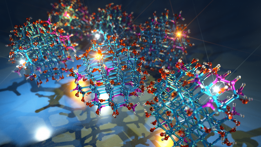
Scientists have developed a nanosystem using “quantum dots” that will help improve visualisation of tumour, paving the way for early treatment.
The nanosystem, which achieves a five-fold increase over existing tumour-specific optical imaging methods, generates bright tumour signals by delivering quantum dots to cancer cells without any toxic effects.
Read also:Gene therapy may treat diabetes and obesity via skin!
According to a research:
- Tumour imaging is an integral part of cancer detection, treatment.
- And tracking the progress of patients after treatment.
- We were able to achieve a tumour-specific contrast index.
- Between five-and 10-fold greater than the general cut-off for optical imaging, which is 2.5.
- The new method utilises quantum dots tiny particles that emit intense fluorescent signals when exposed to light.
- And an “etchant” that eliminates background signals.
- The quantum dots are delivered intravenously and some of them leave the bloodstream and cross membranes, entering cancer cells.
- Fluorescent signals emitted from excess quantum dots that remain in the bloodstream are then made invisible by injecting the etchant.
- The etchant and the quantum dots undergo a “cation exchange”.
- In addition,that occurs when zinc in these are swapped for silver in the etchant.
- The silver-containing quantum dots lose their fluorescent capabilities.
- And because the etchant cannot cross membranes to reach tumour cells,
- Furthermore,the quantum dots that have reached the tumour remain fluorescent.
- Thus, the entire process eliminates background fluorescence while preserving tumour-specific signals.
- Researchers developed the novel method using mice harbouring human breast, prostate and gastric tumours.
Quantum dots were actively delivered to tumours using iRGD, a tumour penetrating peptide that activates a transport pathway that drives the peptide along with bystander molecules in this case fluorescent quantum dots into cancer cells.

